The 19 Muscles Of The Foot - Layers of Sole of Foot _01 | Foot anatomy, Anatomy and ... / Then the wrapped foot is pulled up against the resistance?. Explore the muscles of the foot in this complete guide! This article outlines the basic anatomy of the foot bones. Like the muscles in the rest of the body, it's important to keep the muscles in the feet strong. Interestingly the dorsal foot muscles generally have no insertion at the little toe. Containing 26 bones, 33 joints, 19 muscles and 57 ligaments, it's one of the few pieces of anatomy that can compete with the hand for sheer complexity.
The foot incorporates countless muscles, bones, tendons and ligaments into simple motion and this chart covers them all. Contrary to expectations, the intrinsic foot muscles contribute minimally to supporting the arch of the foot during walking and running. Origin, insertion, innervation and function. The skeleton of the foot is often subdivided, based on functional and clinical 10.16 the short muscles of the right foot from the plantar view. The ultrasound appearances of ankle and foot ligaments, tendons, and nerves are similar to those in other parts of the body.
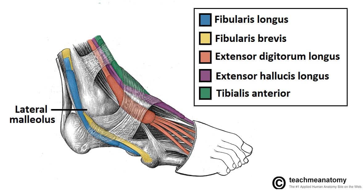
Muscles in the Lateral Compartment of the Leg - TeachMeAnatomy from teachmeanatomy.info The muscles acting on the foot • the muscles acting on the foot can be divided into two distinct groups; This article outlines the basic anatomy of the foot bones. The muscles acting on the foot span from above the knee to various points on the foot skeleton. Maximum isometric force for the main pims is 375 n. The muscles acting on the foot can be divided into two distinct groups; First layer • the first layer of muscles is the most superficial to the sole, and is located immediately underneath the plantar fascia. There are 2 neurovascular planes between the muscle layers of the sole Explore the muscles of the foot in this complete guide!
Neurovascular planes of the sole:
• flexor digiti minimi brevis. Maximum isometric force for the main pims is 375 n. However, these muscles do influence our ability to produce forward propulsion from one stride into the next, highlighting their role in bipedal locomotion. 10.19 (a) pattern of peripheral sensory innervation in the right lower limb. The tendons are thick bands that connect muscles to bones. Supports the longitudinal arch of the foot. Origin, insertion, innervation and function. Can you name the all the muscles of the foot? Which of the following muscles would be strengthened by this exercise; A generous moment arm of these muscles about the midfoot. Layer 3 of the foot. The muscles in the plantar region of the foot may be divided into three groups, in a similar manner to those in the hand. Muscle layers of the sole of the foot.
A generous moment arm of these muscles about the midfoot. To get started, all you need to do is click on the title of the article below that you are most interested in. • this video covers the anatomy of the lumbrical muscles of the foot: Talofibulare anterius (17) lig minimal plantar and dorsal movements and rotation. Barefoot runners make contact with the balls of their feet, the arch of the foot dissipating the energy of the impact safely.
Pin on muscle_foot from i.pinimg.com • flexor digiti minimi brevis. Layer 3 of the foot. Explore the muscles of the foot in this complete guide! Muscles & tendon sheaths of the foot. • fourth layer ( fig. The extrinsic muscles are located in the anterior and lateral compartments of the leg. Supports the longitudinal arch of the foot. Test your knowledge on this science quiz and compare your score to others.
Insertions of the extrinsic foot muscle tendons on the plantar surface of the foot.
Muscles are in the leg, but their tendons function within the foot. There are 29 muscles associated with the human foot. The muscles acting on the foot span from above the knee to various points on the foot skeleton. Their limited impact on posture and movement has led to the broad use of the extensor hallucis brevis and extensor digitorum brevis as muscular sources for tissue grafts. This means that the little toe can only be extended by the extensor digitorum longus muscle only. The muscles in the plantar region of the foot may be divided into three groups, in a similar manner to those in the hand. The foot is an intricate part of the body, consisting of 26 bones, 33 joints, 107 ligaments, and 19 muscles. 4 in each foot, each with 2 heads o: This article outlines the basic anatomy of the foot bones. Foot muscle forces & deformities. • flexor digiti minimi brevis. • this video covers the anatomy of the lumbrical muscles of the foot: • fourth layer ( fig.
The dorsal aponeurosis of the toes supports the effect of the dorsal foot muscles by redirecting the force line of their tendons to. Terms in this set (14). There are 29 muscles associated with the human foot. The muscles in the plantar region of the foot may be divided into three groups, in a similar manner to those in the hand. Origin, insertion, innervation and function.
Muscle: Lower Leg & Foot from cdn.thinglink.me The muscles covered in this article serve various. The dorsal aponeurosis of the toes supports the effect of the dorsal foot muscles by redirecting the force line of their tendons to. The other 19 muscles are referred to as intrinsic muscles of the foot and act only within the foot. There are 2 neurovascular planes between the muscle layers of the sole A generous moment arm of these muscles about the midfoot. There are many ligaments in the foot. There are over two dozen. Flexion of 4 lesser toes at metatarsophalangeal, proximal & distal interphalangeal joints inversion of foot plantar flexion of ankle.
Their limited impact on posture and movement has led to the broad use of the extensor hallucis brevis and extensor digitorum brevis as muscular sources for tissue grafts.
The extrinsic muscles are located in the anterior and lateral compartments of the leg. Talofibulare anterius (17) lig minimal plantar and dorsal movements and rotation. The skeleton of the foot is often subdivided, based on functional and clinical 10.16 the short muscles of the right foot from the plantar view. Flexor hallucis longus tendon transfer to the dorsum of the foot and release of the flexor digitorum longus and brevis tendons at the base of each toe. 26.19 intrinsic muscles of the dorsum right foot, dorsal view. Sides of adjacent metatarsals i: Neurovascular planes of the sole: Ankle ligaments are best examined with the transducer aligned with the long axis of the ligament and the ligament stretched. Barefoot runners make contact with the balls of their feet, the arch of the foot dissipating the energy of the impact safely. The ultrasound appearances of ankle and foot ligaments, tendons, and nerves are similar to those in other parts of the body. Then the wrapped foot is pulled up against the resistance? • flexor digiti minimi brevis. The muscles acting on the foot can be divided into two distinct groups;


0 Komentar